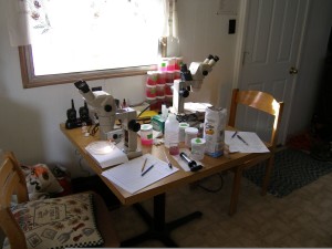
I’ve spent a lot of time in front of a microscope either chasing around a micro invertebrate to analyzing a histological stain for Q/A. During lab courses in school I was always the one person that was good at following around single celled organisms so everyone could get a glimpse at it.
When working at the Desert Studies Center, Zzyzx, CA on the Mohave Tui Chub project, I spent huge amounts of time in front of a stereo dissecting microscope counting zooplankton. It was very tedious work, but a really fun way to explore a world invisible to the naked eye.

At Caris Lifesciences, working in the immunohistochemistry (IHC) lab required immense amounts of microscopy work. We had really nice stereo compound microscopes that every single slide went through. When I was making tissue micro-arrays (TMA) I had to quality control (QC) all kinds of tissue to make sure they would work for use in the TMA. I also spent a lot of time looking at each of our IHC stains to make sure they stained properly and the stainer machines were still staining with the intensity we were looking for. In order for a slide to leave the lab and get in front of a pathologist, it had to go through our internal quality analysis (Q/A) team.
