The staple of the histology lab is microtomy of formalin fixed paraffin embedded (FFPE) blocks. The microtomes and our process at Caris Lifesciences were all validated for 4μ thick sections, so all of my experience is cutting sections that thin. Frozen microtomy uses thicker sections, and some test require thicker sections to work, but we’ve found that 4μ are about one cell thickness. This allows for great visual representation of protein expression in cancer cells after our IHC staining process.
Microtomy can easily be the most frustrating thing to do in the laboratory. It only becomes so if the tissue is processed incorrectly, which a lot of the tissue I’ve sectioned has been poorly processed. There is a special window of just enough water in the tissue. Too much and it cuts poorly, and too little it just turns into powder or hardens to the point of ruining blades. Improperly decalcified bone specimens are also a bear to cut. Usually you just get what you can with those.
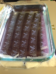
The process we used when doing microtomy is a bath full of heated water and ice blocks. The block goes on the ice first and then the microtome. The microtome chuck is then lined up to the face of the paraffin block and the first initial sections are cut. This is called facing the block. Depending on how that went, you would then either set the block back on the ice, or in the warm water bath.

After the initial facing of the block, you try to get a good ribbon going where each slice connects to the other due to the friction of the tissue and paraffin sliding off the blade. This ribbon is transported very careful to the warm water bath where the paraffin becomes clear. Each segment of the ribbon is teased apart using forceps, and then gently moved and picked up with a glass slide. The slides then go in a drying rack and then into ovens to bake the tissue on the slides.

Once the slides are baked, the tissue has adhered quite well to positively charged glass slides. At this point, the tissue is ready for all the testing it’s going to be enduring. Some processes require a nuclear fast red to make the tumor area pop out, and then the tumor is scraped off the slide under a dissection scope. The powder then runs through an assay to get the DNA/RNA into well plates for molecular analysis. The IHC lab I worked in ran a lot of IHC tests on each case. All of this work would be impossible without the use of microtomy.
I’m familiar with the Leica 2235 and 2255 microtomes. The first is a manual microtome, and the second is an automated one with a foot pedal. I have yet to see anyone use the automation feature on those microtomes. I don’t prefer one over the other, they both cut excellently.


